Multimedia Gallery
Project allows users to explore 3D vertebrate specimens from inside out (Image 7)
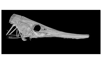
A computed tomography scan showing the skull of a grooved razor fish (Centriscus scutatus) from India. The scan was taken as part of a project to scan vertebrate specimens, then make the data available on an open access website. [Image 7 of 11 related images. See Image 8.]
More about this image
A recent grant by the National Science Foundation (NSF) is supporting the launch of a project that will scan 20,000 vertebrates from the Florida Museum of Natural History's collection using computed tomography (CT) scanning and make these data-rich, 3D images available to researchers, educators, students and the public via the internet.
Called the oVert project -- short for openVertebrate, the project will encompass representative specimens from more than 80% of existing vertebrate genera, and a selection of these will also be scanned with contrast-enhancing stains to characterize soft tissues.
CT scanning takes X-rays of a specimen from every angle, creating thousands of snapshots that a computer stitches together into a detailed 3D visual replica. The image can be virtually dissected, layer by layer, to expose cross sections and internal structures. The scans allow scientists to view a specimen inside and out, including its skeleton, muscles, internal organs, parasites and even its stomach contents, without touching a scalpel.
Images scanned for the project will then be housed in MorphoSource, a public database created by Duke University that scientists, educators, students or the curious can mine for 3D data on their species of interest.
[Research supported by NSF grant DBI 1701714.]
To learn more about this project, see the Florida Musum story New project allows web users to explore 3-D vertebrate specimens from inside out. (Date image taken: 2016-2017; date originally posted to NSF Multimedia Gallery: Dec. 14, 2017)
Credit: Edward Stanley and David Blackburn, Florida Museum of Natural History
Images and other media in the National Science Foundation Multimedia Gallery are available for use in print and electronic material by NSF employees, members of the media, university staff, teachers and the general public. All media in the gallery are intended for personal, educational and nonprofit/non-commercial use only.
Images credited to the National Science Foundation, a federal agency, are in the public domain. The images were created by employees of the United States Government as part of their official duties or prepared by contractors as "works for hire" for NSF. You may freely use NSF-credited images and, at your discretion, credit NSF with a "Courtesy: National Science Foundation" notation.
Additional information about general usage can be found in Conditions.
Also Available:
Download the high-resolution JPG version of the image. (665.3 KB)
Use your mouse to right-click (Mac users may need to Ctrl-click) the link above and choose the option that will save the file or target to your computer.

